PD Dr. Johannes Rheinlaender
Academic Councillor
| Room: | C9 A38 |
| Phone: | +49-7071-29-76031 |
| Fax: | +49-7071-29-5093 |
| E-Mail: |
Key activities
| Atomic force microscopy (AFM) |
| Developments in scanning ion conductance microscopy (SICM) and advanced applications |
| Finite element modeling (FEM) in scanning probe microscopy (SPM) |
| Cell mechanics |
| Biostatistics |
Summary
Understanding the mechanics of the living cell as the smallest building block of life is of major importance in modern life sciences, as cell mechanics turn out to be crucial for many cellular processes in health and disease. For example, cell mechanics are involved in many fundamental cell functions such as adhesion, migration, tissue and organ development, and differentiation. Moreover, numerous diseases are associated with an alteration of the mechanics at the single-cell level. 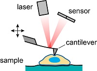 Schematic of AFM setup for live cell imaging. However, many aspects of these processes are not yet fully understood, in large parts because of a lack of quantitative methods to probe the mechanics of living cells at the microscopic scale. Scanning probe microscopes, especially the atomic force microscope (AFM) and the scanning ion conductance microscope (SICM), are ideally suited for this purpose as they allow studying living cells under physiological conditions. The AFM (see schematic right) is a well-established tool for cell imaging, based on bringing a cantilever with a nanometer-sharp tip in contact with the sample. The direct mechanics contact then allows to mechanically probe the sample with even nanometer resolution. Currently, we further develop the AFM for nano-rheological measurements of viscoelastic sample properties. 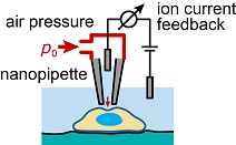 Schematic of SICM setup to The SICM is a less-known scanning probe microscope based on measuring the ion current through a glass nanopipette (see schematic left). Owing to the contact-free feedback principle, it is particularly suited for the imaging of soft and delicate biological samples such as living cells. As many central technical aspects of the SICM are still unknown, we also investigated the image formation process in SICM, for example the resolution and the ion current vs. distance behavior. We furthermore develop methods for mechanically probing the sample with the SICM, for example by inducing a microfluidic flow through the nanopipette locally deforming the sample (see schematic). We thereby combine the contact-free imaging principle of the SICM with quantitative nanomechanical measurements, which is a promising technique for the investigation of the living cell and its cytoskeleton (see images below). 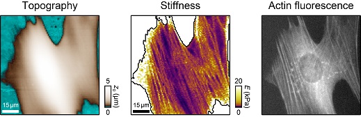 SICM stiffness mapping of a living fibroblast cell with images of topography (left panel) and Due to the complex geometries involved, many scanning-probe-based methods cannot be described analytically. Therefore, I also develop numerical approaches to investigate the performance and to develop new quantitative imaging modes for AFM and SICM. 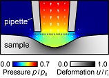 FEM model of fluid flow, pressure, and For example, we used finite element modelling (FEM) to describe the microfluidic flow in SICM stiffness mapping (see model left). |
Curriculum vitae
| 2021 | Habilitation at the Department of Physics, University of Tübingen |
| since 2016 | Academic councillor at the Institute of Applied Physics, University of Tübingen |
| 2016 | Visiting PostDoc at the Department of Physiology, Development and Neuroscience, University of Cambridge, GB |
| 2011-2016 | Research associate at the Institute of Applied Physics, University of Tübingen |
| 2012 | Ph.D. (Physics) |
| 2008-2011 | Research associate at the Institute of Applied Physics, University of Erlangen-Nürnberg |
| 2007 | Diploma in physics |
| 2003-2007 | Studies of physics at the University of Münster |
Awards / Fellowships
| 2018 | Award for Best Study Course in the graduate program 'Biomedical Technologies' |
| 2017 | Award for Best Instructor in the graduate program 'Biomedical Technologies' |
| 2016 | DFG Grant to Support the Initiation of International Collaboration |
| 2015 | Award for Best Instructor in the graduate program 'Biomedical Technologies' |
| 2015 | DGfB Travel Grand EBSA 2015 |